A Medicine Company
When you live with a disease, you depend on medicines to have a chance at a healthier life. That's why we're hard at work making medicines for the people who need them.
A Medicine Company
When you live with a disease, you depend on medicines to have a chance at a healthier life. That's why we're hard at work making medicines for the people who need them.
00:00-00:04
(Metronome ticks)
00:02
Picture of kitchen cupboard with a "CUPS" label.
00:06
(Kettle hisses) The kettle is taken off the stove.
00:09
A woman puts a jacket on and leaves the house.
00:10
A sign behind the woman says, "CHECK W/ DAVID BEFORE LEAVING."
00:12
A close-up of a journal is shown. The word "remember" is written.
00:13
A view from the passenger seat of a car watches a man driving.
00:14-00:15
A husband says, "Oh my god," as he videos his pregnant wife in the mirror.
00:16
Keys are on a key holder on the wall of the house. One key is labeled “HOUSE KEYS ONLY.”
00:17-00:19
A note is seen on the bathroom mirror. It says, "Cancel Dr appointment Mon." The word “Cancel” has been crossed out, and in different handwriting below is written, "Keep it, Hun. I'll drive you."
00:20-00:21
The woman, as a young child, is seen in old home movie-style footage dancing on the beach in a red-skirted bathing suit.
00:22
In the home, an old swimming trophy is seen on a shelf with other awards.
00:22
There is a close-up of a desk calendar with the date of the 24th shown. The message on the date says, "BOOK CLUB CANCELLED THIS WEEK.” Under “BOOK CLUB” is written, but it has been crossed out.
00:23
Many family photos and children's art projects are seen hung up on the refrigerator.
00:24
A close-up of a journal is shown with the words, "I miss them."
00:25-00:26
A phone is seen ringing. Next to the phone is an envelope that's being used as a notepad. It says, "Try Anjali Again." Under that, three different dates are written and have been crossed out.
00:27
As the phone still rings, a family picture is sitting on a side table in the home.
00:28-00:30
The answering machine is heard: "At the tone, please record your message." A woman is seen in the background getting ready for the day. In the foreground, a note is taped to a clock that says, "You haven't upset anyone."
00:32
The answering machine beeps, and old home movie footage of the pregnant woman shown earlier is playing. She is laughing in a garden.
00:34
A close-up of a journal is shown. Words say, "One day at a time."
00:36
A couple is holding hands. The man is wearing a gold wedding band.
00:38
A woman is seen sitting up in bed abruptly while it is still dark outside.
00:40
A light is turned on and a small handwritten sign can be seen on a table that says, "You are home."
00:41
Husband and wife embrace, and the husband asks, "Honey, are you alright?"
00:42-00:50
A clock begins ticking, and as it continues, moments of the woman's life are shown. There are various scenes of her life as she's growing up, and we see her looking at her journal with the words "You are still you” written.
00:51-00:53
The woman sits on her bed, looking at her journal. The text on the screen reads, "People with Alzheimer's take notes to remember."
00:54-00:56
The woman, as a young child, is again seen dancing on the beach in a red-skirted bathing suit. The text on the screen reads: "30 years of research hasn't forgotten them."
00:57-00:60
As the young child continues dancing on the beach, the text on the screen fades, and the Lilly logo appears: "Lilly, A medicine company."
Don't Forget
Innovation takes decades of persistence, but we’re making progress in Alzheimer’s disease. Watch our latest film.

Making an Impact Beyond Medicine
Beyond our medicines, we positively impact society through our work to improve lives and communities around the world.
Our Latest Medicines and Products
Learn more about what's new for patients.
Insulin Affordability
Learn about changes that make it easier for people to get Lilly insulin for $35 or less a month.
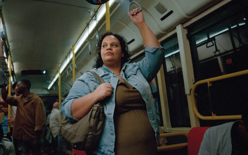
Navigating Obesity: Liz’s Story
Liz’s experience is personal, but it isn’t unique. People living with the disease have many of the same challenges. Now, it’s time to better understand what it’s like.
Driving Innovation for Clinical Trials Access
We’re on the road with a new approach to clinical trials—one that expands access to cutting-edge medicines and treatments.